The lower jaw in non-mammalian vertebrates consists of several bones, including an articular and a dentary, among others, and the lower jaw is joined to the skull by the articular of the lower jaw articulating (and thus the name) with the quadrate of the skull. In the mammal-like reptiles, we see a tendency for quadrate and articular, which lie close to the eardrum, to become smaller and more loosely connected to the skull, while the tooth-bearing dentary becomes enlarged. In some advanced mammal-like reptiles, the jaw joint involves both the quadrate and the squamosal dorsally and the articular and dentary ventrally. Eventually the mammalian joint is solely between the squamosal of the skull and the dentary.
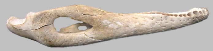
Lower jaw of an American Alligator, showing the sutures between bones making up the jaw. The bone to the far right (showing the alveoli for teeth) is the dentary, which is homologous with the entire lower jaw of mammals. The concavity at the left end of the jaw is part of the articular and forms the connection with the quadrate of the skull.
In non-mammalian tetrapods with functional ears, the only middle-ear ossicle is the stapes (derived evolutionarily from the hyomandibular, a gill-arch-derived bone that braces the upper jaw in fishes. In mammals, the stapes is joined by the incus and malleus (also thought to originally have been derived from ancestral gill arches). These two elements are the old reptilian quadrate and articular, respectively. With loss of the jaw-joint function, they were free to become adapted to other uses, frequent occurrence in evolution.
In mammals, the otic capsule (the structure surrounding the inner ear) is formed by the fusion of centers of ossification which are variable in number; the resultant bone is known as the periotic and generally is buried rather deeply in the skull (the posterior end may reach the surface as the mastoid, however). In early mammals and most marsupials today, the adjacent middle ear (with its incus, malleus, and stapes) had little protection from injury. In most placentals, a bony structure (the auditory bulla, plural bullae) surrounds most of the middle ear. In some, this consists of the tympanic bone, believed from embryological evidence to be the old angular bone of the lower jaw (not to be confused with the angular process of the dentary). In some mammals, a new cartilage bone, the entotympanic, joins the tympanic to form a compound auditory bulla.
If the bulla and periotic fuse with the squamosal, the complex is called the temporal bone.
 Since we're talking about ears, this might be a reasonable time to review
the other function of the inner ear, which is orientation. Three semicircular canals at right angles to each other detect turning motion, while the utriculus and sacculus register the
tilt of the head and linear accelerations. All work via sensory "hairs" (the semicircular canals by stimulating the hair-like structures by movement of a liquid, the utriculus and sacculus by
gelatinous masses containing calcium carbonate, the otoliths, pulling on the structures).
Since we're talking about ears, this might be a reasonable time to review
the other function of the inner ear, which is orientation. Three semicircular canals at right angles to each other detect turning motion, while the utriculus and sacculus register the
tilt of the head and linear accelerations. All work via sensory "hairs" (the semicircular canals by stimulating the hair-like structures by movement of a liquid, the utriculus and sacculus by
gelatinous masses containing calcium carbonate, the otoliths, pulling on the structures).
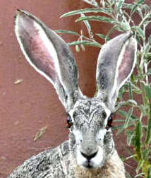
External to the tympanum (ear drum), the auditory canal leads to the opening of the ear (auditory meatus) to the outside;partially surrounding the auditory meatus in most mammals and usually extending dorsally and posteriorly, is a pinna (plural: pinnae). This flap of skin and connective tissue helps guide sound into the ear canal and aids in determining the direction from which the sound is coming.
A Black-tailed Jackrabbit (Lepus californicus) with two large pinnae (one angled partially back) extending from its head. Photograph by Lauri L. Lear.
Last Update: 17 Jan 2008
Centennial Museum and Department of Biological Sciences, The University of Texas at El Paso